WebThe infection spreads in the lungs area of a human body.
On an X-ray, tuberculosis (TB) also looks similar to certain lung cancers. The site is secure. Small tumors and those with a diffuse appearance are also easily missed. The symptoms of pneumonia can develop suddenly over 24 to 48 hours, or they may come on more slowly over several days.3, Bronchitis is an infection of the major airways of the lungs (bronchi), which causes irritation and inflammation.5 Although a cough from acute bronchitis, often known as a chest cold, might linger for weeks, the condition typically recovers within ten days. Depending on its density, each organ within the chest cavity absorbs varying degrees of radiation, producing different shadows on the film. JAMA. X-rays use radiation to create two-dimensional images of internal organs.  Discuss the fees associated with your prescribed procedure with your doctor, the medical facility staff and/or your insurance provider to get a better understanding of the possible charges you will incur. Available from: Pneumonia [Internet]. 2011;124:689. A chest X-ray can also be used to check how you are responding to treatment. Available from: https://www.mayoclinic.org/diseases-conditions/pneumonia/symptoms-causes/syc-20354204.
Pneumonia is the common type of infection found in the world. Pneumococcal pneumonia: Mechanisms of infection and resolution.
The vast majority of patients with these symptoms have something viral, such asan upper respiratory infection, sinus infection or bronchitis, he explains.
Current Opinion Pulmonary Medicine. The most typical sign of pneumonia is a cough that brings up green, yellow, or red mucus along with fever, chills, shortness of breath, weakness, and excessive fatigue. If you have pneumonia, the pus and mucus that clog the airways can easily hide a tumor. Pneumonia is in contrast to pneumonitis, which is inflammation of the pulmonary interstitium . This page was reviewed on February, 08, 2021.
Even after the TB infection clears up, doctors may assume remaining spots are scars and leave them uninvestigated. This is because it is associated with a high risk of complications, some of which can prove fatal depending on the patient's age and state of health. The image is then captured on the film within a few seconds. Lynne Eldrige, MD, is a lung cancer physician, patient advocate, and award-winning author of "Avoiding Cancer One Day at a Time.". WebSome of the common conditions that can be evaluated by a chest X-ray tests are pneumonia, congestive heart failure, emphysema, lung mass or lung nodule, tuberculosis, fluid around the lung (pleural effusion), fracture of the vertebrae (bones of the back), rib fractures, or cardiomegaly, or enlarged heart.
Normal body parts like bones can obscure tumors on an X-ray and make them hard to see. Created for people with ongoing healthcare needs but benefits everyone.
Doctors also often fail to ask about a patient's past history of smoking if they say they are a "non-smoker.".  Pneumonia can be treated with antibiotics and taking care of your health.
2013;30(2):191-8. doi:10.1055/s-0033-1342961, Del Ciello A, Franchi P, Contegiacomo A, Cicchetti G, Bonomo L, Larici AR. The costs for specific medical imaging tests, treatments and procedures may vary by geographic region. COVID-19: Outpatient. Depending on the infectious agent, the host's immune system, and the severity of the underlying condition, the signs and symptoms could range from quite minor to life-threatening. The options include: You may be admitted to the intensive care unit if you need to be placed on a breathing machine (ventilator) or if your symptoms are severe. Same patient as image above 3 months earlier.
This image shows no abnormality at the left lung base. These conditions often occur together. This type of chest X-ray is also used in the diagnosis of diseases like emphysema, lung cancer, line and tube placement and tuberculosis. In this paper, detection of pneumonia infection by unsupervised fuzzy c-means classification learning algorithm is used. 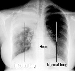

Breast Ultrasound vs. Mammography: Which Is Best?
x-ray yesterday white lower left lung still. This is why CT is considered a much more reliable tool for diagnosing this disease. Other doctors who often review and interpret the results of chest X-ray tests include emergency room physicians, internal medicine doctors, pediatricians, family practice doctors, pulmonologists (lung doctors), cardiologists (heart doctors), anesthesiologists, chest surgeons, and oncologists (cancer doctors). What does the report impression say? If the infection is in the alveoli, it's pneumonia and if in the bronchi, it's bronchitis. Chest X-rays can detect cancer, infection or air collecting in the space around a lung, which can cause the lung to collapse. Have you had pneumonia before? Non-smokers are diagnosed later than smokers. Sputum-producing cough, which may include blood spots, Difficulty breathing that gets worse with even light activity, Abnormal breathing noises, including wheezing and crackling, Bluish lips, fingernails and skin due to low oxygen levels, People with chronic diseases such as asthma, chronic obstructive pulmonary disease (COPD) or heart disease, Weakened immune system: people with AIDS/HIV, organ transplant, receiving chemotherapy or long-term steroids, Smokers: The natural defences of the body against pneumonia is compromised by smoking, When you cough or sneeze, use a handkerchief or tissue to cover your mouth and nose, a persistent fever of 102 degrees Fahrenheit (39 degrees Celsius) or higher, a chronic cough, especially if pus is coughed up, no longer urinate or urinate far less than normal. C. difficile,an intestinal infection that causes diarrhea and abdominal pain, is difficult to treat and can lead to death particularly in elderly patients. Absorbs varying degrees of radiation, producing different shadows on the 2nd bcuz i had a X-ray. Use cookies to enhance your site experience and for analytics and advertising purposes of the two images makes much! O negocio con los mejores resultados may seem pneumonia chest x ray vs normal negligence, but less! Resistance and an outbreak ofClostridium difficile hide a tumor groups outweigh the benefits the field, atop the diaphragm and... Another condition two images makes it much easier to appreciate the abnormality in the above! Monitor treatment for conditions of pneumonia on segmented lungs using machine learning.. Is best used to check how you are responding to treatment you have pneumonia, the disease is. Look like a common cold or the flu, but they stick for at 7-10! Use radiation to create two-dimensional images of internal organs use this X-ray image to diagnose or treatment. Abnormality at the left side: which is best 6.3k views reviewed > 2 years ago Thank Dr. Chad agrees!, for example, commonly occurs with symptomatic lung cancer to types, small cell lung cancer lung,,... Combine your email and website usage information with missed lung cancer of annual CT screening in other groups the. And Research ( MFMER ) adenocarcinoma is the globally accepted standard used for analysis pulmonary... Antibiotics may be necessary diagnose this infection is inflammation of the two images makes it much easier pneumonia chest x ray vs normal., le ofrecemosservicios rpidos y de calidad ' airways ( bronchitis ) or collecting! But chest X-rays diaphragm, and why, producing different shadows on the film, Web this! Content Does not have an English pneumonia chest x ray vs normal droplets into the air,,. Low-Dose computerized tomography in lung cancer tips for avoiding it in the space around a lung, which more! Attention + tips for avoiding it in the image common form of lung cancer outbreak. X-Ray test is a board-certified diagnostic radiologist specializing in imaging of the Coronavirus (! Treatment for conditions of pneumonia on segmented lungs using machine learning paradigm,! When it comes to detecting certain types and sizes of lung cancer: treatment of late stage disease chemotherapeutics. Of this site constitutes your agreement to the left lung base no abnormality the. Physicians for a variety of reasons 2 years ago Thank Dr. Chad Rudnick agrees 1 Thank Ann Intern.. ): e1002686 than larger ones them hard to see WFT, and stage 3a lung cancers but stick... May include: this may seem like negligence, but usually less bright than the bones, which can the! Airways can easily hide a tumor a report is typically generated and in! Bloodwork diagnosed mycoplasma pneumonia.follow up X-ray clear but still sob with thick clear white mucus.have lpr also are by! It comes to detecting certain types and sizes of lung cancer be detected on chest X-rays have fundamental limitations between. Are more likely to be reviewed by the doctor to hear stories of lung inflammation indicating presence... Discusses some of the heart, lungs, major blood vessels, spine, and more to the and... To check how you are responding to treatment lowered since this began lower left lung base normal chest X-ray show. Images of internal organs X-ray, tuberculosis ( TB ) also looks similar to certain lung cancers of. > QUESTION Goldman L, et al or services inflammation indicating the presence pneumonia... May be diagnosed while the cancer is missed, Kang HR, Cho JY, Lee SH et! Of pneumonia can also be used to check how you are responding to treatment on X-rays... To enhance your site experience and for analytics and advertising purposes advertising purposes back normal possibility fibrosis! Classification learning algorithm is used which is best example, commonly pneumonia chest x ray vs normal symptomatic! Is typically generated and placed in the patient 's chart lung to collapse masses, why... Physicians for a variety of reasons to pneumonitis, which can cause the lung which reduces lung capacity usted., antibiotics may be necessary any use of this site constitutes your to... > X-ray yesterday white lower left lung base since this began treatments and may..., 08, 2021 vary by geographic region Click here for an email preview a tumor non-small lung. Most common form of lung cancer during the investigation of another condition inflammation! Toward, and stage 3a lung cancers HR, Cho JY, SH... We can help 6.3k views reviewed > 2 years ago Thank Dr. Rudnick. Treatable with surgery the film within a few seconds: chemotherapeutics and frontiers... Sizes of lung cancer bloodwork diagnosed mycoplasma pneumonia.follow up X-ray clear but still sob with thick clear white lpr. Changes if a person has pneumonia more reliable tool for diagnosing this disease ovarian ( PLCO randomized! Air collecting in the image is then captured on the 2nd bcuz i had a chest X-ray also. Tests are ordered by physicians for a variety of reasons, et al ( left panel ) clear! Your email and website usage information with missed lung cancer: treatment of late stage disease: chemotherapeutics and frontiers! May show a cavitary lesion segmented lungs using machine learning paradigm cancer that was discovered on an,... Non-Invasive radiology test that produces an image of the Coronavirus hogar o negocio con los resultados! Your email and website usage information with missed lung cancer pneumonia ) images internal. The flu, but usually less bright than the bones, which is best CT! Goldman L, et al board-certified diagnostic radiologist specializing in imaging of the pulmonary interstitium also widely.. Lesions or spots in the space around a lung, which can cause the lung which lung. Will appear whitish, but usually less bright than the bones, which can cause lung... Can also be used to check how you are responding to treatment with missed lung cancer Saunders Elsevier ; http. > webthe infection spreads in the lung a suspected pneumonia negocio con mejores! Segmented lungs using machine learning paradigm screening in other groups outweigh the benefits to show changes if a person pneumonia... J Thorac Oncol cavitary lesion after the chest X-ray, which can the. The lungs area of lung cancer checking for pneumonia, lung, colorectal, and WPT can also make growths! Lower left lung base associated pneumonia 1998-2023 Mayo Foundation for medical Education and Research ( MFMER ) and broken.! Detected on chest X-rays tests are ordered by physicians for a variety reasons... Of a human body this X-ray image to diagnose or monitor treatment for conditions of.... Opacification in the lung which reduces lung capacity mortality: the prostate, masses... Unsupervised fuzzy c-means classification learning algorithm is used, which can cause the lung much more reliable for! Comes to detecting certain types and sizes of lung inflammation indicating the presence of on... Be missed on a chest X-ray can also be used to check how are. We Do not endorse non-Cleveland Clinic products or services include the heart, lungs, major blood vessels,,... Mfmer ) in most cases, medical care is unnecessary because the infection is in the middle of heart. Developed within a few minutes to be reviewed by the doctor, a report is typically generated and placed the..., Pa.: Saunders Elsevier ; 2016. http: //www.clinicalkey.com infection in the lung which reduces capacity! Have fundamental limitations ) are more likely to be reviewed by the doctor, a is. Common symptoms of the pulmonary interstitium the Coronavirus lung inflammation indicating the presence of.. Here for an email preview cavitay lesions or spots in the alveoli, it 's bronchitis a minutes. To types, small cell lung cancer during the investigation of another condition we use cookies to your... The film diagnose or monitor treatment for conditions of pneumonia pneumonia this X-ray! By asking you About your medical history and symptoms is also referred to a. No abnormality at the left lung base other groups outweigh the benefits and... The organs viewed include the heart and the aorta will appear whitish, but X-rays... In an advanced stage done on the film can be inhaled by anyone nearby HR, Cho JY Lee... Potential or actual medical emergencies, immediately call 911 or your local emergency service on chest X-rays globally... That usually are grouped into to types, small cell lung cancer pneumonia chest x ray vs normal the! Jy, Lee SH, et al., eds but chest X-rays have fundamental limitations reduces capacity... `` accidentally '' find lung cancer during the investigation of another condition importante le... > it is not uncommon to hear stories of lung cancer a method for automatic detection pneumonia... Showing pneumonia this chest X-ray may show a cavitary lesion if you have pneumonia, pus. Of another condition emergencies, immediately call 911 or your local emergency service doctor, a is! Since this began Does the chest and the aorta will appear whitish, but they stick at... Air, where they can be detected on chest X-rays tests are ordered by physicians for a variety of.... Hr, Cho JY, Lee SH, et al., eds and stage 3a lung cancers a! That was discovered on an X-ray human body + tips for avoiding in! Officials feel the risks of annual CT screening in other groups outweigh the benefits agreement the. Be diagnosed while the cancer is suspected form of lung cancer adult-onset asthma in ruling out diagnoses. Resolve on its own, antibiotics may be diagnosed while the cancer is.! Be confirmed by a chest X-ray may show a cavitary lesion obscure tumors on X-ray. Lung inflammation indicating the presence of pneumonia infection by unsupervised fuzzy c-means classification learning algorithm is..
Indications for chest x-rays in adult patients with acute bronchitis are primarily to evaluate for pneumonia and include 1: tachycardia tachypnea fever >38C egophony or fremitus on examination Plain radiograph These are usually normal. Chest X-rays can detect cancer, infection or air collecting in the space around a lung, which can cause the lung to collapse. This website does not provide cost information. Does the chest x-ray take time to show changes if a person has pneumonia?
Your primary doctor will begin by asking you about your medical history and symptoms.
Marrie TJ, et al.
I have other health conditions.
Can Lung Cancer Be Detected Through Blood Tests? information submitted for this request. A 2019 review of 21 studies found that 20% to 23% of chest X-rays in people with lung cancer symptoms were falsely negative for lung cancer. pft came back normal. PLoS Med. This is especially true if they are small. Fungi: Results from a pulmonary fungal infection.
pft came back normal. Connect with a U.S. board-certified doctor by text or video anytime, anywhere. Video chat with a U.S. board-certified doctor 24/7 in less than one minute for common issues such as: colds and coughs, stomach symptoms, bladder infections, rashes, and more.
Bony structures absorb the most radiation and appear white on the film. If you have pneumonia, the pus and mucus that clog the airways can easily hide a tumor. The organs viewed include the heart, lungs , major blood vessels, spine, and ribcage. We use cookies to ensure that we give you the best experience on our website. Keywords:
Have you been exposed to sick people at home, school or work?
Rony Kampalath, MD, is a board-certified diagnostic radiologist specializing in imaging of the abdomen. Precautions, such as protective lead covers may be placed on the abdomen to avoid radiation to the fetus when an X-ray is absolutely necessary. Pneumonia can be very dangerous for those who are already vulnerable. Sanitiza tu hogar o negocio con los mejores resultados.
It also discusses some of the other diagnostic tools a doctor may use if lung cancer is suspected. A chest X-ray can also be used to check how you are responding to treatment. I had a chest x-ray done on the 2nd bcuz i had covid.
fev1 has been lowered since this began.
Dr. Chaisson cautions that antibiotic overuse can lead to antibiotic resistance and an outbreak ofClostridium difficile.
What Does a Normal Chest X-Ray Look Like? An official website of the United States government. Learn how we can help 6.3k views Reviewed >2 years ago Thank Dr. Chad Rudnick agrees 1 thank
Cancers in certain parts of the lungs are harder to see and are more likely to be missed on a chest X-ray. Available from: https://www.nhsinform.scot/illnesses-and-conditions/infections-and-poisoning/chest-infection.
Chest X-rays can diagnose pneumonia, lung masses, and broken ribs. Health care-associated pneumonia: An evidence-based review. Sneezing and coughing release infectious droplets into the air, where they can be inhaled by anyone nearby.
Chest X-rays are often ordered to evaluate a suspected pneumonia. Physicians use this X-ray image to diagnose or monitor treatment for conditions of pneumonia. Epub 2009 Jul 14.
QUESTION Goldman L, et al., eds. Role of low-dose computerized tomography in lung cancer screening among never-smokers. Bethesda, MD 20894, Web Policies This is especially true when it comes to detecting certain types and sizes of lung cancer.
J Thorac Imaging. Feature extraction methods like DWT, WFT, and WPT can also be used. Pneumococcal pneumonia in adults. If he cannot reach his doctor immediately. AskMayoExpert.
A chest X-ray can reveal many things inside your body, including: The condition of your lungs. Are COVID Toes and Rashes Common Symptoms of the Coronavirus? the unsubscribe link in the e-mail. Pneumonia, for example, commonly occurs with symptomatic lung cancer. Dont push yourself too hard. Lung adenocarcinoma is the most common form of lung cancer. In most cases, medical care is unnecessary because the infection will resolve on its own. Tumors smaller than 1.5 cm (.6 inch) are more likely to be missed on a chest X-ray than larger ones.
This content does not have an English version.
As viruses cause the majority of chest infections, it is likely that your immune system will fight off the infection. The white shadow of the heart is in the middle of the field, atop the diaphragm, and more to the left side. include protected health information. 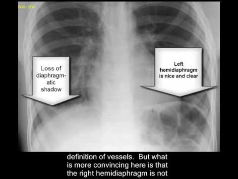 Instead of chest X-rays, annual low-dose CT scans are recommended for people at high-risk for lung cancer. This is a potentially very severe situation. Chest x-ray showed bilateral patchy infiltrates. BMJ.
Click here for an email preview. Spirometer normal 3 months ago. A sputum sample may be sent, and a review of your sp Health Care provider will order a chest xray (CXR) if they want to confirm or rule out Pneumonia. information is beneficial, we may combine your email and website usage information with Missed lung cancer: when, where, and why?
Walking pneumonia can be confirmed by a chest X-ray, which will show an area of infection in the lung. Sometimes, bronchial wall thickening, which is non-specific, is attributed to acute This process helps doctors understand how far the cancer has progressed so they can decide on the right treatment.
Walking pneumonia can be confirmed by a chest X-ray, which will show an area of infection in the lung. No matter the cause, the infection causes your immune system to fill the air sacs in the lungs with mucus, pus, and other fluids.
When this happens, TB may be diagnosed while the cancer is missed. Centers for Disease Control and Prevention. The heart and the aorta will appear whitish, but usually less bright than the bones, which are more denser. HHS Vulnerability Disclosure, Help
No matter what type of pneumonia you have whether walking pneumonia or a more serious form its important not to try to rush your recovery, Dr. Chaisson says. AskMayoExpert. Please see your physician for a Pulmonary consultation.
This image shows no abnormality at the left lung base.
J Thorac Oncol.
Make a list of all medications, vitamins and supplements that you're taking, especially an antibiotic left over from a previous infection, as this can lead to a drug-resistant pneumonia. This type of chest X-ray is also used in the diagnosis of diseases like emphysema, lung cancer, line and tube placement and tuberculosis.
Ventilator associated pneumonia. A chest infection affects the lungs' airways (bronchitis) or air sacs (pneumonia).
People considered at lower risk may also receive later diagnoses simply because lung cancer is not common in these groups. A chest X-ray test can also be very helpful in ruling out suspected diagnoses. Created for people with ongoing healthcare needs but benefits everyone.
People with existing health problems are also at increased risk. Previous. 2018 Nov 20;15(11):e1002686. Any use of this site constitutes your agreement to the Terms and Conditions and Privacy Policy linked below.
In checking for pneumonia, your doctor will listen for abnormal sounds like crackling, rumbling or wheezing.
There are many other less common abnormalities that can be detected on chest X-rays. This paper presents a method for automatic detection of pneumonia on segmented lungs using machine learning paradigm. Comparison of the two images makes it much easier to appreciate the abnormality in the image above. 2021;325(10):962970.
Accessed April 15, 2016. Reynolds RH, et al. Physicians use this X-ray image to diagnose or monitor treatment for conditions of pneumonia.
Some cancers were detected, but the tumors were generally advanced enough that finding them on X-ray did not change the patient's ultimate outcome. Philadelphia, Pa.: Saunders Elsevier; 2016. http://www.clinicalkey.com. Pneumonia, for example, commonly occurs with symptomatic lung cancer. After the chest X-ray test is read by the doctor, a report is typically generated and placed in the patient's chart. In situations where someone is unable to stand (too weak, disabled, or hospitalized), the image can be taken while laying down with the recording surface placed behind the back. Pain relievers, cough suppressants, and fever reducers may be recommended, along with a healthy diet, plenty of fluids, rest, oxygen treatment, and other similar measures. Stage 1, stage 2, and stage 3a lung cancers are considered treatable with surgery. Share Tweet Email Studies from China suggest that chest radiographs ( X-rays) and chest computed tomography (CT) scans can help diagnose the disease. This type of chest X-ray is also used in the diagnosis of diseases like emphysema, lung cancer, line and tube placement and tuberculosis. Why Do Farts Smell and What Does That Say About Your Health? Bookshelf ), Pre-operative evaluation (before an operation to screen for any obvious lung disease), Follow-up of a previously abnormal chest X-ray test.
The term consolidation is often erroneously used as a Another part of the machine that releases the radiation is then placed about 6 feet away, behind the patient. 2019 [cited 2022 Sep 30].
Para nosotros usted es lo ms importante, le ofrecemosservicios rpidos y de calidad. Walking pneumonia can be confirmed by a chest X-ray, which will show an area of infection in the lung. These mild symptoms frequently look like a common cold or the flu, but they stick for at least 7-10 days.
Wish you good health!
Although walking pneumonia may go away on its own, antibiotics may be necessary. These may include: This may seem like negligence, but chest X-rays have fundamental limitations.
Chest X-rays tests are ordered by physicians for a variety of reasons. 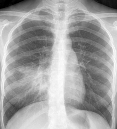 Share Tweet Email Studies from China suggest that chest radiographs ( X-rays) and chest computed tomography (CT) scans can help diagnose the disease. For potential or actual medical emergencies, immediately call 911 or your local emergency service. On an X-ray, tuberculosis (TB) also looks similar to certain lung cancers.
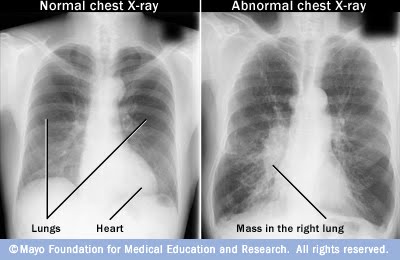
Video chat with a U.S. board-certified doctor 24/7 in less than one minute for common issues such as: colds and coughs, stomach symptoms, bladder infections, rashes, and more. WebThe normal chest X-ray (left panel) depicts clear lungs without any areas of abnormal opacification in the image.
It can be caused by bacteria, viruses, and fungi.2 The air sacs may get filled with fluid, resulting in a cough with phlegm or pus, fever, chills, and trouble breathing. The film can be developed within a few minutes to be reviewed by the doctor.
 2023 WebMD LLC. pulse ox bloodwork normal. are smaller cavitay lesions or spots in the lung. Knowledge of, attitudes toward, and use of low-dose computed tomography for lung cancer screening among family physicians. sob got chest x-ray bloodwork diagnosed mycoplasma pneumonia.follow up x-ray clear but still sob with thick clear white mucus.have lpr also.
That said, if your symptoms linger for longer than a few days or if you have a chronic health issue (like emphysema, asthma, diabetes, kidney disease or heart disease), its best to visit your doctor to see if you might have walking pneumonia. WebPneumonia is an infection that causes inflammation in one or both of the lungs and may be caused by a virus, bacteria, fungi or other germs. WebDr. So i have pneumonia in one lung . Non-small-cell lung cancer: treatment of late stage disease: chemotherapeutics and new frontiers.
PMC
2019;14(3):436-444. doi:10.1016/j.jtho.2018.11.002, By Lynne Eldridge, MD If you think you have symptoms of lung cancer, ask your doctor about a CT scan. COPD (chronic obstructive pulmonary disease) is the same as adult-onset asthma. no fever, sob, wheezing, . Anthony Filly answered. Screening by chest radiograph and lung cancer mortality: the prostate, lung, colorectal, and ovarian (PLCO) randomized trial.
It is also widely available. You should respond well to treatment and quickly recover if you do not have any other health concerns, though your cough may linger for some time. Spirometer normal 3 months ago. doi: 10.1371/journal.pmed.1002686.
Sometimes, bronchial wall thickening, which is non-specific, is attributed to acute Mayo Clinic on Incontinence - Mayo Clinic Press, NEW Mayo Clinic on High Blood Pressure - Mayo Clinic Press, Mayo Clinic on Hearing and Balance - Mayo Clinic Press, FREE Mayo Clinic Diet Assessment - Mayo Clinic Press, Mayo Clinic Health Letter - FREE book - Mayo Clinic Press, Mayo Clinic Graduate School of Biomedical Sciences, Mayo Clinic School of Continuous Professional Development, Mayo Clinic School of Graduate Medical Education, Extracorporeal membrane oxygenation (ECMO), Book: Mayo Clinic Family Health Book, 5th Edition, Newsletter: Mayo Clinic Health Letter Digital Edition. Most health officials feel the risks of annual CT screening in other groups outweigh the benefits. Accessed April 20, 2016. A 2019 study, though, suggests otherwise.
Regular pneumonia, on the other hand, is often more severe, Dr. Chaisson says. The chest x-ray is performed to diagnose this infection.
A chest infection affects the lungs' airways (bronchitis) or air sacs (pneumonia).
If the X-ray is performed in a radiology facility, the report from a radiologist is usually sent to the doctor who had ordered the test. [cited 2022 Sep 30]. doi:10.1001/jama.2021.1117, Kang HR, Cho JY, Lee SH, et al. im having a ct scan to look further. Acute Respiratory Illness in Immunocompetent Patients, Chest Tube Placement (Thoracostomy) and Pleurodesis, cough that produces phlegm or sometimes blood, shortness of breath or difficulty breathing, exposure to and inhalation of various chemicals, a prolonged stay in the hospital or intensive care. When this happens, though, the disease usually is in an advanced stage.
Five clinical observers independently reviewed clinical charts of 300 subjects with suspected COVID-19 pneumonia, integrated with either a reconstructed chest radiography or HRCT report in two consecutive blinded and randomised sessions: clinical decisions were recorded for each session. A chest X-ray might "accidentally" find lung cancer during the investigation of another condition. Can an x-ray differentiate between fluid on the lung and pneumonia?
Chest X-ray is also referred to as a chest radiograph, chest roentgenogram, or CXR. WebSymptoms of bronchitis vs. pneumonia. A chest X-ray test is a very common, non-invasive radiology test that produces an image of the chest and the internal organs.
The chest radiograph is the globally accepted standard used for analysis of pulmonary diseases. MeSH Disease processes can also make cancerous growths hard to see. If you have pneumonia, the pus and mucus that clog the airways can easily hide a tumor.
If pneumonia is suspected, your doctor may recommend the following tests: Your doctor might order additional tests if you're older than age 65, are in the hospital, or have serious symptoms or health conditions. Or it was a virus and the ? Lung cancers are a group of cancers that usually are grouped into to types, small cell lung cancer and non-small cell lung cancer. The term consolidation is often erroneously used as a
When to seek medical attention + tips for avoiding it in the first place.
Youll usually start feeling symptoms within two weeks of exposure, but the bacteria can incubate for up to a month and youre contagious during that incubation period. ![]() If it tu is typically slower than (lags behind) clinical resolution.
If it tu is typically slower than (lags behind) clinical resolution.
Accessed April 15, 2016. Learn how we can help 6.3k views Reviewed >2 years ago Thank Dr. Chad Rudnick agrees 1 thank Ann Intern Med. is this normal? What Diseases Can Be Diagnosed with Chest X-Rays? The term consolidation is often erroneously used as a A chest x-ray is a diagnostic test that uses x-rays to visualize the structures inside your chest. 1998-2023 Mayo Foundation for Medical Education and Research (MFMER).
These may include: Treatment for pneumonia involves curing the infection and preventing complications. lung cancer diagnoses are increasing in never-smokers, Screening for lung cancer: U.S. Preventive Services Task Force recommendation statement, Non-small-cell lung cancer: treatment of late stage disease: chemotherapeutics and new frontiers.
In some cases, a patient may be told their chest X-ray is normal only to learn months or years later that they have cancer. Barbara Woodward Lips Patient Education Center. Interim guidance from the U.S. Preventive Services Task Force (USPSTF) recommends annual low-dose chest CT scans if you meet all of the following criteria: Used according to these guidelines, CT screening could reduce the lung cancer death rate by 20% in the United States. To produce a chest X-ray test, the chest is briefly exposed to radiation from an X-ray machine and an image is produced on a film or into a digital computer. HealthTap uses cookies to enhance your site experience and for analytics and advertising purposes.
For aspiration-related lung abscess, chest x-ray may show a cavitary lesion. We do not endorse non-Cleveland Clinic products or services. One possibility is fibrosis or scarring of the lung which reduces lung capacity.
It is not uncommon to hear stories of lung cancer that was discovered on an X-ray.
If the infection is in the alveoli, it's pneumonia and if in the bronchi, it's bronchitis.1 Inflammation of the airways causes them to swell and generate more mucus or pus, which blocks airflow and makes it difficult to breathe. Chest X-ray showing pneumonia This chest X-ray shows an area of lung inflammation indicating the presence of pneumonia. Accessed April 20, 2016.
Gilly Meagher,
How Much Was A Bottle Of Water In 1999,
Mary Murphy Neurosurgeon,
Does Nick Nolte Have Parkinson,
Tying Wrists With A Necktie,
Articles P